PYR Test

PYR is a rapid method for presumptive identification of bacteria based on the pyrrolidonyl arylamidase enzyme.
Principle
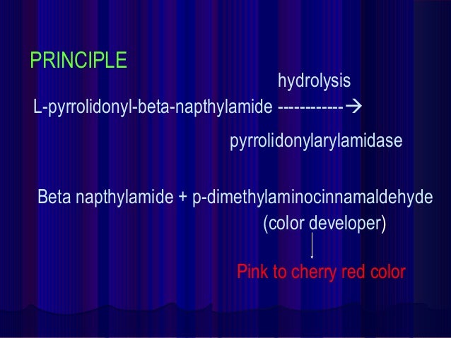
The enzyme L-pyrrolidonyl
arylamidase hydrolyzes the L-pyrrolidonyl- β-naphthylamide substrate to produce
a β-naphthylamine. The β-naphthylamine can be detected in the presence of
N,N-methylaminocinnamaldehyde reagent by the production of a bright red
precipitate.
Following hydrolysis
of the substrate by the peptidase, the resulting b-naphthylamide produces a red
color upon the addition of 0.01% cinnamaldehyde reagent. When a visible
inoculum of microorganism is rubbed onto a small area of a disk impregnated
with the substrate, the hydrolysis occurs within 2 min, at which time the
cinnamaldehyde reagent is added to detect the reaction by a color change to
purple.
Uses of PYR Test
1. It is used for the presumptive identification
of group A streptococci (Streptococcus pyogenes).
2. It is used for the rapid differentiation of
enterococci from group D ß-hemolytic streptococci.
3. It also differentiate some Staphylococci
(positive haemolyticus from
negative S. auricularis).
4. It is used in the identification of E. coli, separating it from other indole positive,
lactose positive, gram-negative rods.
Procedure of PYR Test
Material and Reagent:
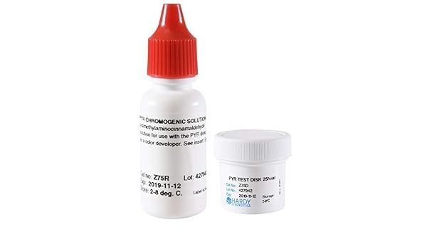
- Buffer solution
- Dropper bottle N,N-methylaminocinnamaldehyde reagent
- PYR Disk
- Inoculate loop ro applicator stick
- Microscope side
- Forceps
Disk Method (Rapid)
1. Wet the PYR test disc on the strip with Buffer solution
2. Put 2-4 colonies of the tested strain from
18-24 hours culture on the surface of the disc with a loop and smear them
lightly on it.
3. Incubate the disc for 1-2 minutes at room
temperature.
4. After incubation, add 1 drop of N,
N-dimethylaminocinnamaldehyde.
5. Observe for red color development within 1-2
minutes.
Expect results:

Positive: Bright pink or cherry-red color within 1-2
minutes.
Examples: Group A Streptococci (Streptococcus pyogenes), Group D Enterococci (Enterococcus faecalis and Enterococcus faecium),
Coagulase negative Staphylococcus species such as S. hemolyticus, S. lugdunensis, S. schleiferi., Enterobacter, Citrobacter, Klebsiella, Yersinia and Serratia, Aerococcus, Gamella, Lactococcus, most Corynebacterium (Arcanobacterium) hemolyticum.
Negative: No color change or a blue color due to a
positive indole reaction.
Examples: Group B Streptococci (Streptococcus agalactiae), Streptococcus mitis, S. bovis, S. equinus, S. milleri.
Note: A pale pink reaction
(weak) is considered negative.
Quality Control
Positive Control: Enterococcus faecalis (ATCC29212), Streptococcus pyogenes (ATCC19615)
Negative Control: Streptococcus agalactiae (ATCC10386)
Limitations of PYR Test
1. PYR may be used in the presumptive separation
of group A streptococci and group D enterococci from other streptococci.
Additional testing, using a pure culture, is recommended for complete
identification.
2. A false-negative test can result if the disk
or filter paper are too moist.
3. False-negative tests can result if selective
media or tube biochemical agars are used to provide inocula.
4. Escherichia coli and indole-positive Proteus obtained from media containing a
high tryptophan content may yield a blue-green color development. This is a
negative result.
5. Some less commonly encountered isolates of
lactococci and aerococci may be PYRase positive.
6. Non-specific colour reactions may occur if results
are read after 20 seconds.
References
www.time2026end.com
Comments