
Microscopic examination of urine

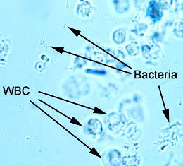
Microscopic examination of urine expressing microscopically observed leucocytes, erythrocytes, and casts in urine, both with centrifugation,

Testing Image result for microscopic examination of urine

Specimen:
Urine patient
Equipment:
tube, centrifuge,timer,pipette transfer,microscope side, cover slip,microscopy
Testing:

  1. Urine take in tube 3/4 of tube
  2. Take to centrifuge 3000rmp/1mn at 5 minutes
  3. Decant out of above liquid 
  4. Pipette transfer sucks sediment take on microscope side and cover slip
  5. Examination with microscopic
Result:


Image result for microscopic examination of urine
Image result for microscopic examination of urineurine color chart #nursingschool #nurse #RN #study #inspiration #school #tips
Waxy and granular casts in urine.
Reference:
www.time2026end.com
Laboratory book livre de lavoratorie 1th edition 1999
https://www.google.com/url?sa=i&url=http%3A%2F%2Fe-learning.studmed.unibe.ch%2FUroSurf_EN%2Ftheory%2Fsedimethods2.html%3Furosurf%7Ctheory%7Csediment%7Csedimethods%7C2&psig=AOvVaw0KIQHg49jHVyHFdvza_3TL&ust=1582250323839000&source=images&cd=vfe&ved=0CAMQjB1qFwoTCNDK8fmD3-cCFQAAAAAdAAAAABAg

Comments

Popular posts from this blog

Mueller Hinton Agar

Mueller Hinton Agar (MHA)       The major use of Mueller Hinton Agar is for antimicrobial susceptibility testing. Composition of MHA Ingredients   Beef Extract                                  2.00 gm Acid Hydrolysate of Casein         17.50 gm Starch                                           1.50 gm Agar                                             17.00 gm Distilled Water                             1000 ml Final pH 7.3 ± 0.1 at 25ºC Why Mueller Hinton agar is used for antibiotic susceptibility testing? 1.     It is a non-selective, non-differential medium. This means that almost all organisms plated on here will grow. 2.     It contains starch. Starch is known to absorb toxins released from bacteria, so that they cannot interfere with the  antibiotics . It also mediates the rate of diffusion of the antibiotics through the agar. 3.     It is a loose agar. This allows for better diffusion of the antibiotics than most other plates. A bette

Simmons Citrate Test

Simmons Citrate Reaction It  used to differentiate among the Gram-Negative bacilli in the family Enterobacteriaceae. Principle  Citrate agar is used to test an organism’s ability to utilize citrate as a source of energy. The medium contains  citrate  as the sole  carbon source  and  inorganic ammonium salts (NH4H2PO4)  as the sole source of  nitrogen . Bacteria that can grow on this medium produce an enzyme,  citrate-permease , capable of converting  citrate  to  pyruvate .  Pyruvate  can then enter the organism’s metabolic cycle for the production of  energy . Growth is indicative of utilization of citrate, an intermediate metabolite in the  Krebs cycle . When the bacteria metabolize  citrate , the  ammonium salts  are broken down to  ammonia , which increases  alkalinity . The shift in pH turns the  bromthymol blue  indicator in the medium from  green to blue  above pH 7.6. Christensen  developed an alternative citrate test medium that does not require the organism

Urease Test

Urease Test The urease test is used to determine the ability of an organism to split urea, through the production of the enzyme urease. Principle  Urea  is the product of decarboxylation of  amino acids . Hydrolysis of  urea  produces  ammonia  and  CO2 . The formation of  ammonia  alkalinizes the medium, and the pH shift is detected by the color change of  phenol red  from  light orange  at pH 6.8 to  magenta (pink)  at pH 8.1. Rapid urease-positive organisms turn the entire medium  pink  within 24 hours. Weakly positive organisms may take several days, and negative organisms produce  no color change  or  yellow  as a result of  acid production . Uses  1.     This test is used to differentiate organisms based on their ability to hydrolyze urea with the enzyme urease. 2.     This test can be used as part of the identification of several genera and species of Enterobacteriaceae, including  Proteus, Klebsiella ,