MacConkey Agar

MacConkey Agar


MacConkey agar is a selective and differential media used for the isolation and differentiation of non-fastidious gram-negative rods, particularly members of the family Enterobacteriaceae .

Composition


MacConkey agar is used for the isolation of gram-negative enteric bacteria and the differentiation of lactose fermenting from lactose non-fermenting gram-negative bacteria. Pancreatic digest of gelatin and peptones (meat and casein) provide the essential nutrients, vitamins and nitrogenous factors required for growth of microorganisms. Lactose monohydrate is the fermentable source of carbohydrate. The selective action of this medium is attributed to crystal violet and bile salts, which are inhibitory to most species of gram-positive bacteria. Sodium chloride maintains the osmotic balance in the medium. Neutral red is a pH indicator that turns red at a pH below 6.8 and is colorless at any pH greater than 6.8. Agar is the solidifying agent.

Uses of MacConkey Agar
1.    MacConkey agar is used for the isolation of gram-negative enteric bacteria.
2.    It is used in the differentiation of lactose fermenting from lactose non-fermenting gram-negative bacteria.
3.    It is used for the isolation of coliforms and intestinal pathogens in water, dairy products and biological specimens.
Preparation 
1.    Suspend 49.53 grams of dehydrated medium in 1000 ml purified/distilled water.
2.    Heat to boiling to dissolve the medium completely.
3.    Sterilize by autoclaving at 15 lbs pressure (121°C) for 15 minutes. 
4.    Cool to 45-50°C.
5.    Mix well before pouring into sterile Petri plates.
Prepare culture
The specimen streaking on macconkey agar.Incubation 37c 18-24h

Image result for mannitol salt agar strin
Result Interpretation

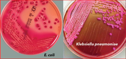
Lactose fermenting strains grow as red or pink and may be surrounded by a zone of acid precipitated bile. The red colour is due to production of acid from lactose, absorption of neutral red and a subsequent colour change of the dye when the pH of medium falls below 6.8.

Lactose non-fermenting strains, such as Shigella and Salmonella are colourless and transparent and typically do not alter appearance of the medium. Yersinia enterocolitica may appear as small, non-lactose fermenting colonies after incubation at room temperature.

References

Comments

Popular posts from this blog

Microscopic examination of urine

Microscopic examination of urine expressing microscopically observed leucocytes, erythrocytes, and casts in urine, both with centrifugation, Testing  Specimen: Urine patient Equipment: tube, centrifuge,timer,pipette transfer,microscope side, cover slip,microscopy Testing: Urine take in tube 3/4 of tube Take to centrifuge 3000rmp/1mn at 5 minutes Decant out of above liquid  Pipette transfer sucks sediment take on microscope side and cover slip Examination with microscopic Result: Reference: www.time2026end.com Laboratory book livre de lavoratorie 1th edition 1999 https://www.google.com/url?sa=i&url=http%3A%2F%2Fe-learning.studmed.unibe.ch%2FUroSurf_EN%2Ftheory%2Fsedimethods2.html%3Furosurf%7Ctheory%7Csediment%7Csedimethods%7C2&psig=AOvVaw0KIQHg49jHVyHFdvza_3TL&ust=1582250323839000&source=images&cd=vfe&ved=0CAMQjB1qFwoTCNDK8fmD3-cCFQAAAAAdAAAAABAg

Triple Sugar Iron Test

The Triple Sugar Iron (TSI) Test  To determine the ability of an organism to ferment glucose, lactose, and sucrose, and their ability to produce hydrogen sulfide. Principle The Triple Sugar Iron (TSI) test is a microbiological test named for its ability to test a microorganism’s ability to ferment sugars and to produce hydrogen sulfide. An agar slant of a special medium with multiple sugars constituting a pH-sensitive dye (phenol red), 1% lactose, 1% sucrose, 0.1% glucose, as well as sodium thiosulfate and ferrous sulfate or ferrous ammonium sulfate is used for carrying out the test.  . Due to the building of acid during fermentation, the pH falls. The acid base indicator Phenol red. is incorporated for detecting carbohydrate fermentation that is indicated by the change in color of the carbohydrate medium from orange red to yellow in the pres...

Urease Test

Urease Test The urease test is used to determine the ability of an organism to split urea, through the production of the enzyme urease. Principle  Urea  is the product of decarboxylation of  amino acids . Hydrolysis of  urea  produces  ammonia  and  CO2 . The formation of  ammonia  alkalinizes the medium, and the pH shift is detected by the color change of  phenol red  from  light orange  at pH 6.8 to  magenta (pink)  at pH 8.1. Rapid urease-positive organisms turn the entire medium  pink  within 24 hours. Weakly positive organisms may take several days, and negative organisms produce  no color change  or  yellow  as a result of  acid production . Uses  1.     This test is used to differentiate organisms based on their ability to hydrolyze urea with the enzyme...